Our Office
-
Room 416, 77 Cheongam-ro, Nam-gu, Pohang, Gyeongbuk, 37673, S.Korea
Ultrasound/Photoacoustic peripheral vascular imaging device
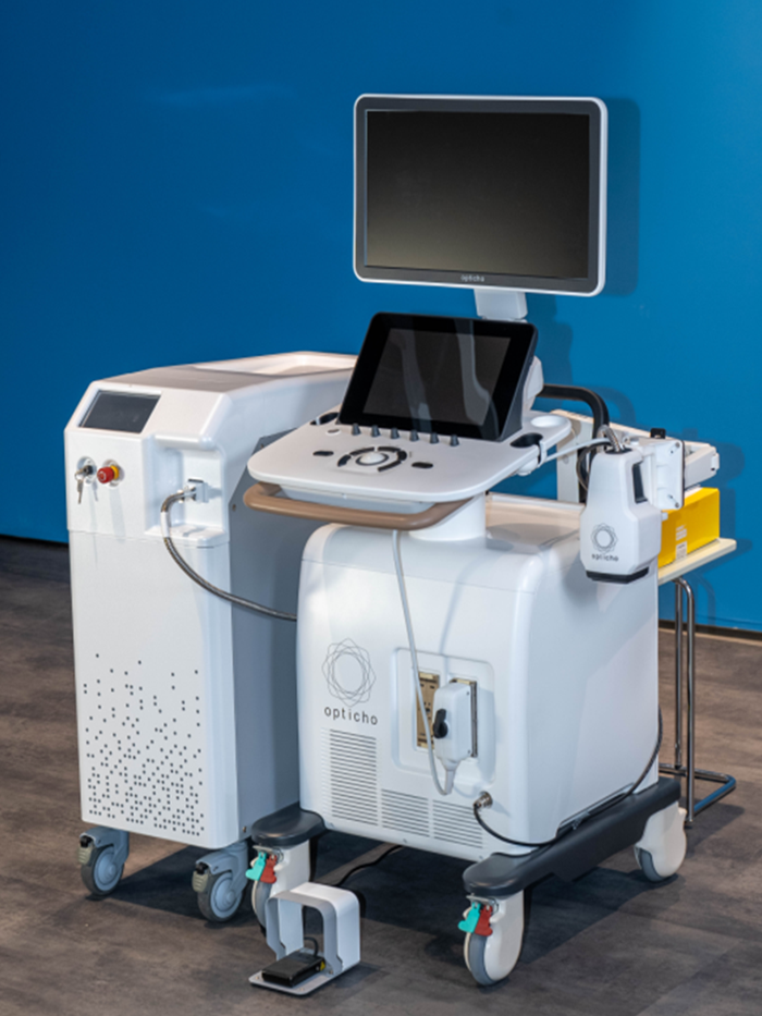
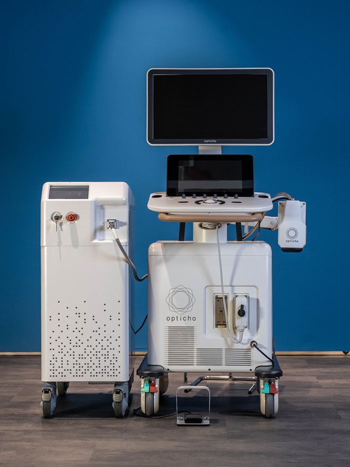
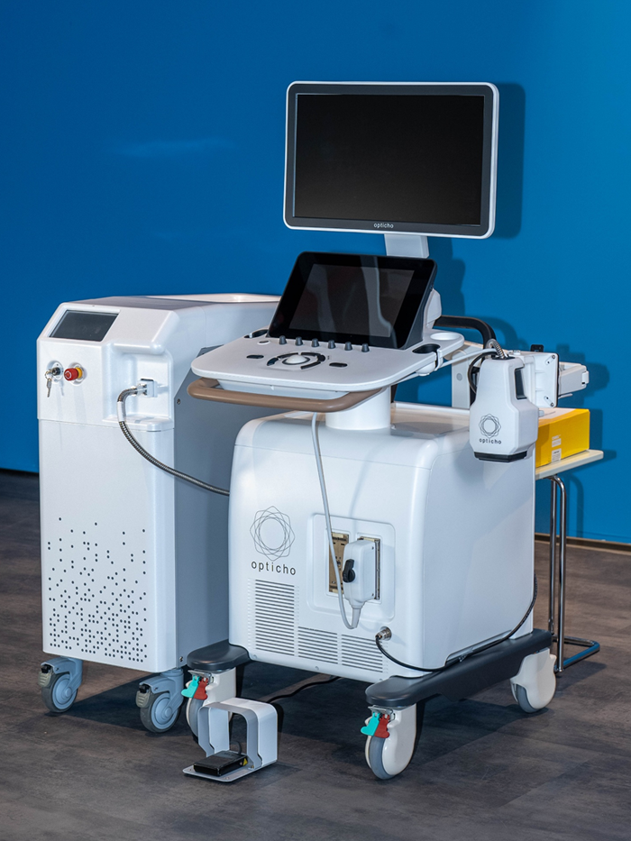
The Perigio medical imaging device visualizes peripheral blood vessels, providing enhanced detail and functional insight. By combining conventional clinical ultrasound with advanced photoacoustic imaging Perigio captures both structural and functional information, including vascular morphology and oxy-/deoxyhemoglobin signals in peripheral microvessels.
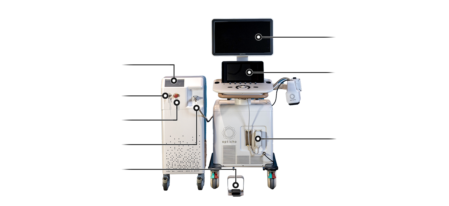
The system integrates a medical-grade laser and an ultrasound imaging module in a safety-compliant design to acquire photoacoustic signals generated by hemoglobin. All core components have been subjected to formal safety evaluations by authorized assessment institutes in the Republic of Korea, in compliance with international standards for medical electrical equipment—including IEC 60601-1 (General Requirements), IEC 60601-1-2 (Electromagnetic Compatibility), IEC 60601-2-22 (Laser Equipment), and IEC 60601-2-37 (Ultrasound Imaging Equipment). The laser delivers light to the body via an optical fiber bundle, while the ultrasound probe detects photoacoustic waves generated by hemoglobin. These signals are processed by dedicated software to reconstruct and visualize vascular structures.
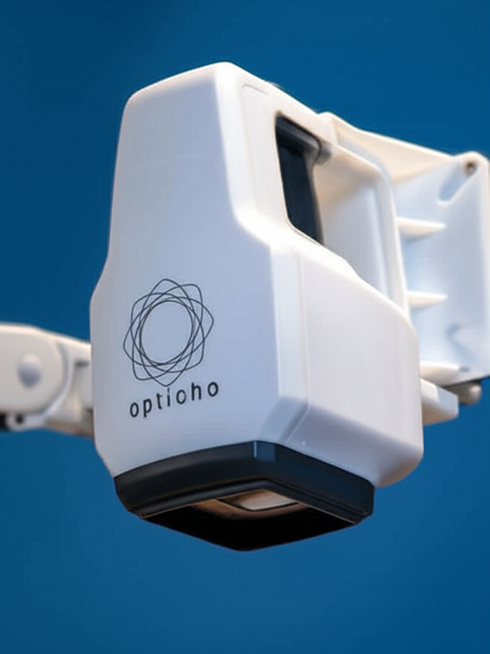
A compact handheld scanner enables volumetric 3D vascular imaging in a user-centric, automated workflow. The scanner integrates an ultrasound probe, optical fiber bundle, and motorized linear actuator in a compact device optimized for one-handed use. It supports efficient acquisition across a wide field of view. Scanning and 3D image reconstruction are automatically performed by the built-in software, activated via a simple foot switch.
Diagnosis and monitoring of peripheral vascular diseases
Diagnosis and monitoring of thyroid cancers, breast cancers, and prostate cancers
Imaging and monitoring of tissue perfusion, vascular status, and wound healing
Evaluation of new drug efficacy
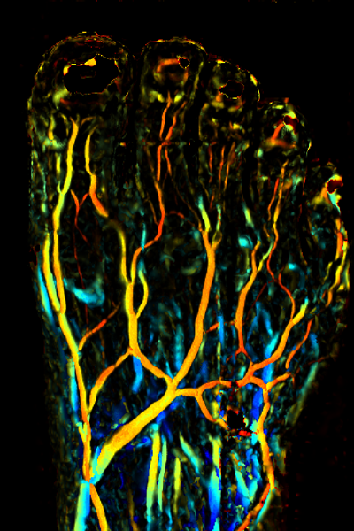
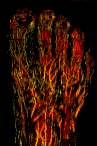
3D microvasculature: Visualization of micro-vessels in extremities of the human body
No contrast agents: Hemoglobin itself is an excellent signal source and daily monitoring available
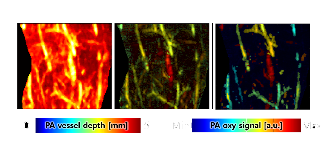
Blood oxygenation: Visualization of photoacoustic oxygenation signal intensity
Vessel pulsation: Ultrasound-guided identification of arterial pulsation with corresponding photoacoustic signal visualization
User-friendly operation: Automatic scan and image acquisition by holding a scanner and pressing a foot switch
Real-time software interface
| Laser | Wavelength | 650 nm and 1064 nm |
|---|---|---|
| Pulse energy | 50 mJ @ 650 nm, 100 mJ @ 1064 nm | |
| Pulse width | 10 ns | |
| Pulse repetition rate | 1 to 10 Hz | |
| Ultrasound | Frequency | 8 MHz |
| Bandwidth | 50% | |
| Image | Field of view | 40 × 40 mm² |
| Spatial resolution | 1 mm |
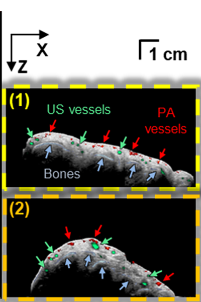
In vivo photoacoustic/ultrasound peripheral angiography of humans
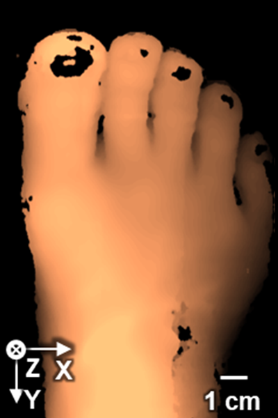
(a) US foot (depth)
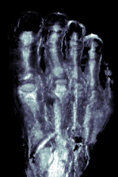
(b) US bone (MIP)
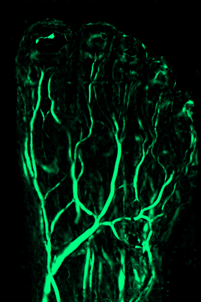
(c) US vessel (MIP)
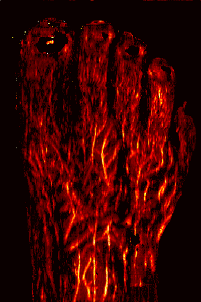
(d) PA vessel (MAP)
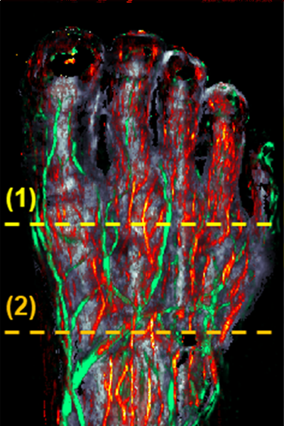
(e) US bone + US/PA vessel
(f) US B-mode + US bone + US/PA vessel
(g) US vessel (depth)
(h) PA vessel (depth)
W. Choi, et al., Radiology, 303(2), 467-473 (2022)
Fusion of light & sound creating new value
© 2024 Opticho. All rights reserved.